
Normal ECG Reading Explained: Essential Insights Everyone Needs to Know
Getting an ECG can feel a bit confusing if you’re not sure what all those spikes and dips mean. But understanding a normal ECG reading doesn’t have to be complicated. It’s just a way for doctors to check how your heart is working by looking at its electrical activity. Whether you’re getting one for a routine check-up or because of symptoms, knowing what’s considered normal can help ease some of the stress. This guide breaks it down in simple terms, so you’ll know what your doctor is looking for, and what it means when they say everything looks fine.
Understanding the Basics of an ECG
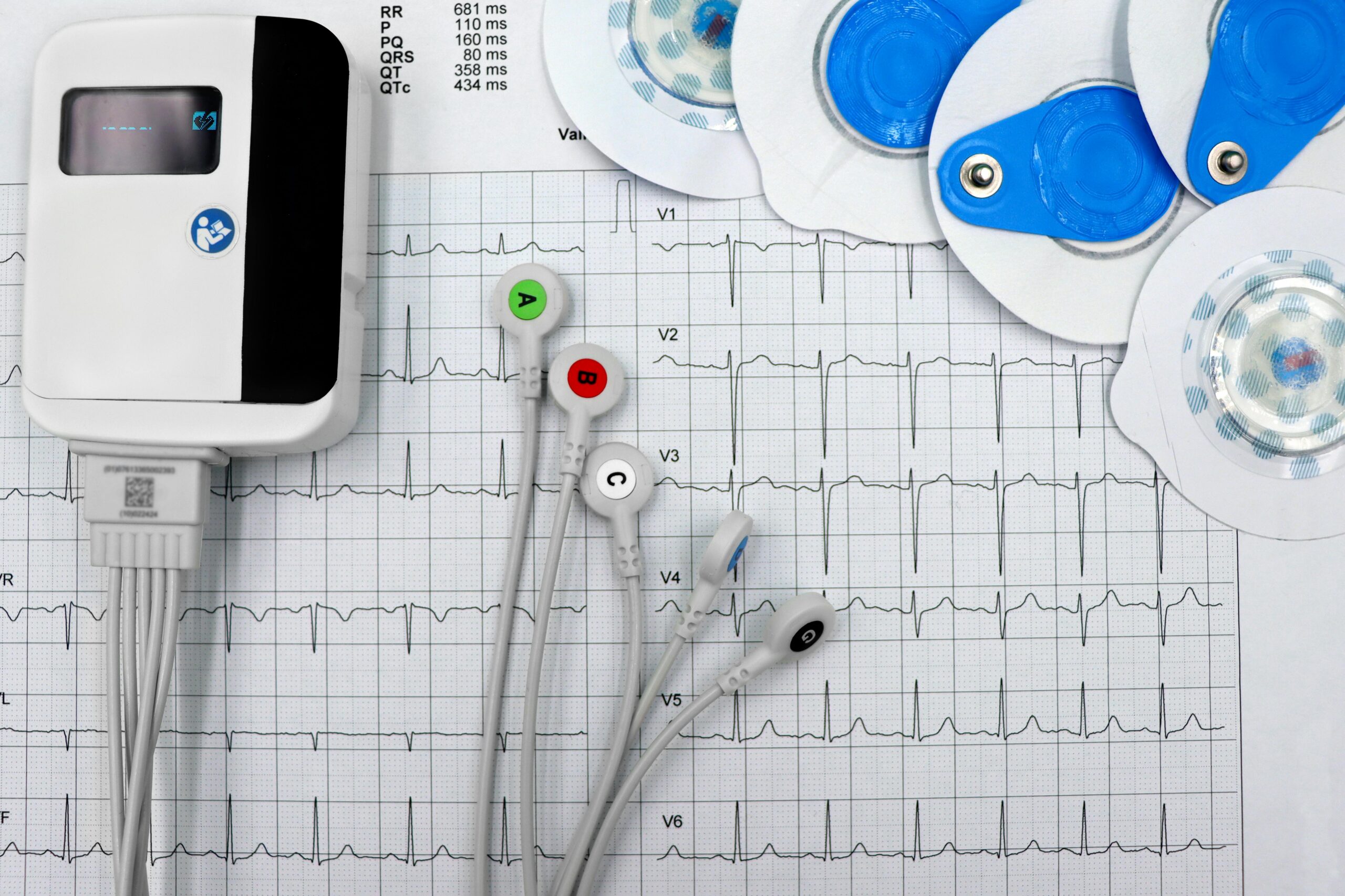
An electrocardiogram, or ECG, is a test that tracks the heart’s electrical signals. It shows how fast your heart beats and whether its rhythm is steady. Small sensors called electrodes stick to the skin on your chest, arms, and legs. These connect to a machine that records patterns made by your heartbeat.
Each time the heart pumps blood, it creates an electric signal. This signal moves through different parts of the heart in a set path. The ECG captures this movement and displays it as lines on a screen or paper. These lines include waves called P waves, QRS complexes, and T waves.
The P wave shows when the top chambers of the heart send blood to the lower ones. The QRS complex reflects how the lower chambers push blood out to your body or lungs. The T wave shows when these chambers reset before starting again.
Doctors read these patterns to see if they match what’s expected from a normal ECG reading. If something looks off, like extra beats, skipped signals, or unusual timing, it could point to issues like irregular rhythms or blocked arteries.
Even though an ECG only takes a few minutes, it gives helpful details about how your heart works at that moment. It’s often used during checkups or before surgeries just to make sure everything looks right.
Learning what each part means helps people understand their results better when they talk with their doctor later on. Gaining this knowledge also makes future tests feel less confusing since you already know what those spikes and dips represent on the graph.
You don’t need any special training to recognise basic patterns once someone explains them clearly. That makes it easier for anyone trying to follow along with their own health updates over time without guessing what every number means during appointments.
Key Components of a Normal ECG Reading
A standard ECG reading shows a simple pattern that repeats with every heartbeat. Each part of the pattern tells something about how the heart is working. The first part to look at is the P wave. This small bump happens when the upper parts of the heart, called atria, send out an electrical signal to start a beat. It shows that the signal begins in the right place and moves through the atria correctly.
Next comes a group of waves known as the QRS complex. These three points show how electricity moves through the lower chambers, or ventricles. The QRS complex is usually quick and sharp because these chambers pump blood out fast and hard. If this section looks off, it might mean there’s a problem with how electricity flows through those areas.
After that comes the T wave. This part shows that the ventricles reset after pumping blood so they can get ready for another beat. A healthy T wave helps confirm that this recovery step happens as it should.
Each piece: the P wave, QRS complex, and T wave, has its own timing too. Doctors check how long each one takes and look at spaces between them to make sure everything runs on schedule.
All these waves together create a full picture of one heartbeat’s electrical activity. By checking if these parts appear in order and within normal limits, experts can tell if someone’s heart rhythm works as expected.
Understanding what each section means helps spot anything unusual early on. When all these elements fall into place without delay or changes in shape, it points toward proper heart function based on what’s seen in a normal ECG reading.
Common Variations That Are Still Considered Normal
Not all ECGs that look different from the textbook examples mean something is wrong. People’s hearts can show a wide range of patterns that still fall within what doctors see as safe. These differences often depend on things like age, activity level, or even certain medicines.
For example, athletes often show slower heart rates on their ECGs. This isn’t bad, it just means their hearts pump more efficiently. A fit person might have a heart rate below 60 beats per minute and still be completely fine. That kind of result might look off at first glance but actually shows strong heart function.
Older adults may also have changes in their ECGs that aren’t dangerous. As people get older, some parts of the electrical signal slow down slightly. This can lead to small shifts in timing between waves or intervals on the graph. These changes don’t always point to illness; they’re just part of getting older.
Some medications also affect how an ECG looks. Drugs for blood pressure or mental health conditions can change the shape or length of certain waves without causing harm. Doctors know how to read these shifts and take medications into account when reviewing results.
Even stress or sleep habits can play a role in temporary changes to your heart’s rhythm on an ECG test. So if you’re tired, anxious, or dehydrated during the test, it might cause slight differences that aren’t serious.
A normal ecg reading doesn’t look exactly the same for everyone because many factors influence how your heart behaves day-to-day. Knowing this helps people avoid panic when they hear terms like “abnormal” from a quick scan of results, context matters a lot with these tests. Talking with your doctor about any unique features in your reading is always helpful before jumping to conclusions about what’s normal for you personally.
When to Seek Medical Advice Based on ECG Results
A normal ECG reading can be helpful, but it doesn’t always mean everything is fine. The ECG looks at your heart’s rhythm and electrical signals. It gives important data, but it doesn’t tell the full story on its own.
Sometimes, people still feel unwell even if their ECG appears okay. Things like chest pain, dizziness, or shortness of breath shouldn’t be ignored. These signs may point to problems that an ECG alone can’t catch. Your body might be giving clues that something deeper is going on.
Let’s say someone has tightness in the chest or gets tired easily when walking up stairs. Even if their test results come back looking fine, those symptoms deserve a closer look. A doctor might suggest more tests like bloodwork or imaging scans to check for other issues.
It’s also common for an ECG to miss brief or hidden irregularities in the heartbeat. Some conditions don’t show up unless they happen during the test itself. That’s why doctors often ask about how you’re feeling overall and not just what shows up on paper.
If you’ve had heart-related symptoms before – or have risk factors like high blood pressure, it’s smart to talk with a healthcare provider even after a normal result. They may want to monitor things over time or run follow-up exams just to stay cautious.
Always listen to what your body tells you and don’t rely only on one test result for peace of mind. If something feels off, it’s better to get checked than wait too long without answers.
Making Sense of What Your Heart Is Telling You
Now that you’ve got a clearer picture of what readings from a normal ECG look like, it’s easier to understand how this simple test offers valuable insight into your heart’s health. From recognising the basic components to knowing which variations can still be considered normal, being informed helps take the mystery out of ECG results. While a normal ECG reading is usually reassuring, it’s always smart to check with a healthcare provider if something feels off. Staying curious and aware is key—because when it comes to your heart, knowledge really is power.